New Preprint: Structural Diversity and Essential Functions of Zebrafish Left-Right Organizer Cilia

by Heidi Hehnly in ,


We are excited to share our new preprint on bioRxiv, Functionally Essential and Structurally Diverse: Insights into the Zebrafish Left-Right Organizer’s Cilia via Optogenetic IFT88 Perturbation and Volume Electron Microscopy . This work was led by Favour Ononiwu (Hehnly lab Graduate student) with contributions from Melissa Mikolaj (NCI- Narayan Lab), Christopher Dell (NCI-Narayan Lab), Abdalla Wael Shamil (Hehnly lab undergraduate), Kedar Narayan (NCI), and Heidi Hehnly.

Why this study matters

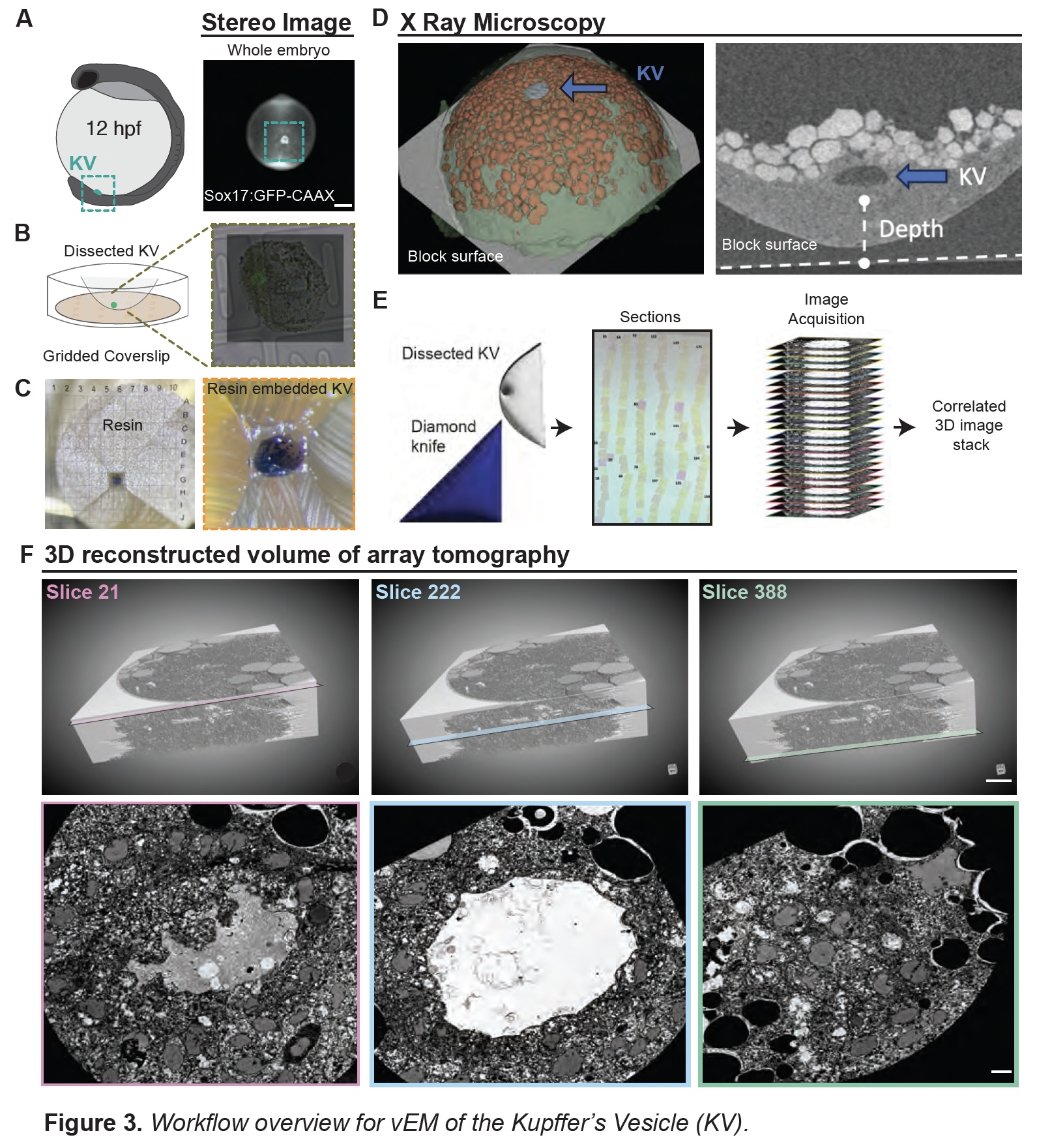
During vertebrate embryogenesis, the left-right organizer (LRO) generates asymmetric fluid flow that initiates left-right body patterning. In zebrafish, this role is carried out by a transient epithelial organ known as Kupffer’s Vesicle (KV). The cilia within KV have long been known to generate flow, but their structural heterogeneity and contribution to KV morphogenesis have remained unclear.

Our approach

We combined two powerful tools:

  • Optogenetics: Using a newly engineered sox17:Cry2-GFP zebrafish line, we clustered the intraflagellar transport protein IFT88 in KV progenitors via blue-light activation. This perturbation impaired ciliogenesis and disrupted lumen formation, establishing a direct role for cilia in KV morphogenesis.

  • Volume electron microscopy (vEM): We generated the first high-resolution 3D ultrastructural map of a mature KV, enabling unprecedented analysis of ciliary architecture across the tissue.

Key findings

  • Only ~70% of cilia retained both mother and daughter centrioles, suggesting that centriole elimination may occur during KV development.

  • Among centrioles, distal appendages (34%), subdistal appendages (92%), and rootlet fibers (5%) were present in highly variable patterns, revealing remarkable structural diversity.

  • Cilia were frequently associated with membrane-bound vesicles, including ciliary-associated vesicles (CaVs) and dense vesicles (CaDVs), with distinct spatial distributions across the KV.

Broader implications

Our findings uncover previously unrecognized complexity in LRO organization. The structural specialization of KV cilia suggests that they may contribute not only to generating flow but also to organizing the architecture of the organ itself. This work adds to a growing appreciation that cilia are not uniform organelles, but instead exhibit context-specific diversity that underpins their function.

Next steps

We anticipate that these insights will inform broader studies of ciliary specialization across tissues and their role in developmental disorders linked to left-right asymmetry.

👉 Read the full preprint here.


New Publication: Dynamic Forces Sculpt Organ Shape in Zebrafish Development

by Heidi Hehnly in ,


We are excited to share our new collaborative paper with the Manning and Amack labs, published in PNAS. This work addresses a fundamental question in developmental biology: how do cells and tissues achieve the precise shapes required for organ function?

Why this matters

Many studies have focused on how cell-intrinsic properties—like signaling pathways or cytoskeletal dynamics—contribute to tissue shape. But development is more than just cells behaving individually; it is also about how tissues as a whole generate and respond to forces. Recent theoretical work suggests that embryonic tissues exist near a “jamming” transition, meaning they can flow very slowly but still transmit large forces over long timescales. These dynamic forces, though often overlooked, have been hypothesized by Manning and Amack groups to play a powerful role in shaping organs.

Our focus: Kupffer’s vesicle

To test this idea, Amack and Manning labs turned to Kupffer’s vesicle (KV), a transient, ciliated organ in zebrafish embryos that our lab also loves to examine. KV plays a crucial role in establishing left-right asymmetry during development, making it an ideal model to study how tissues generate and respond to mechanical forces.

What we did

This project combined mathematical modeling, live imaging, and in vivo perturbations to test whether dynamic forces generated by tissue movements sculpt KV shape. The last part with in vivo perturbations, is where our group played an important role first with Mike Bates (a postbac in our lab and then Manager of the Blatt Imaging Center) and then with Yiling Lan (a graduate student in the lab).

  • Modeling predicted that slow tissue flows during embryogenesis could apply significant stresses to KV, driving its morphological changes.

  • Laser ablation experiments, performed in our lab (by Mike and Lan), were critical to test these predictions. By precisely severing tissue connections in the embryo, we altered force transmission and directly observed the resulting effects on KV shape. The outcomes matched the model predictions, providing strong evidence that dynamic forces are a key driver of organ morphology.

The bigger picture

The collaborative findings show that self-generated dynamic forces sculpt organ shape during development. Because many developmental processes occur on slow timescales, this principle likely applies broadly beyond zebrafish KV. This work opens the door to exploring how tissues harness dynamic mechanical forces across diverse developmental contexts.

We are thrilled to have contributed to this collaborative effort—particularly by performing the ablation experiments—and to see how interdisciplinary approaches combining modeling, physics, and cell biology can shed new light on fundamental developmental mechanisms.

📄 Read the full paper here


Congrats to Hehnly Lab Seniors!

by Heidi Hehnly in , ,


Congrats to Alison Gerety who received a distinction in Biology, was a Renee Crown Honors student, and won an Outstanding Achievement in Biology award for her thesis on “Characterization of Vimentin in Zebrafish Lateral Line Development”!

Congrats to Abdalla W. Shamil who was awarded both Academic and Research Achievements, and was a Renee Crown Honors student!

We can’t wait to see all that you go on to do!

Abdalla, Heidi, and Alison before the Biology Award Ceremony.

Alison at her poster on her Thesis project.


BioArt Talk with Adam Zaretsky and Pop-Up Exhibit by Aksiniya Peycheva Recap

by Heidi Hehnly in , , ,


Last Friday, the Honors Program hosted a compelling BioArt talk by artist and educator Adam Zaretsky in the Lundgren Room. Known for his provocative work at the intersection of art and biology, Zaretsky captivated the audience with an engaging presentation that challenged traditional boundaries between scientific inquiry and artistic expression.

Following the talk, attendees gathered in the second-floor lunchroom of the Life Sciences Complex for refreshments and continued conversation. The event also featured a pop-up exhibit by Aksiniya Peycheva titled Trauma Mapping, which offered a powerful exploration of embodied memory and healing through mixed media.

It was a lively and thought-provoking afternoon of interdisciplinary exchange, drawing students and faculty from across campus.